Homonymous Hemianopia Associated with COVID-19 Vaccination
Homonymous Hemianopia After COVID-19 Vaccination
Vol. 21 No. 1 : 2026 (542-546)

Abstract
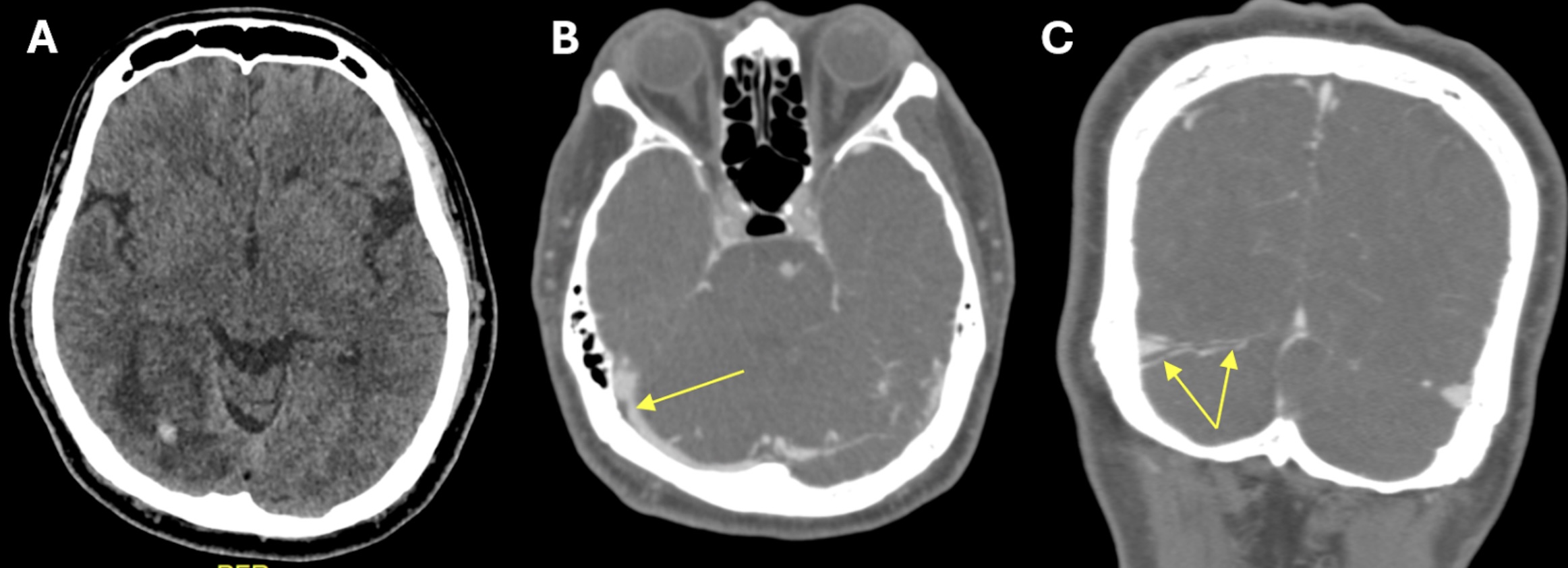
The current COVID-19 pandemic has prompted the development of vaccines to control the spread and severity of SARS-CoV-2. However, there are growing reports of different types of strokes, including ischemic and hemorrhagic strokes, as well as cerebral venous sinus thrombosis (CVST), following COVID-19 vaccination. We presented a case involving a 55-year-old male with a history of ischemic heart disease who exhibited an atypical presentation of visual field impairment one week following Sinovac-CoronaVac COVID-19 vaccination. The patient also experienced mild headache and vomiting for two days preceding the onset of visual symptoms. Right eye’s best-corrected visual acuity was 6/24, and left 6/15. A visual field confrontation test indicated left homonymous hemianopia. An urgent CT venography of the brain documented right transverse sinus thrombosis with occipital haemorrhagic infarct. A nasopharyngeal swab tested negative for SARS-CoV-2, and baseline blood investigations were within normal ranges. The patient was given one week of low-molecular-weight heparin, followed by a three-month course of oral anticoagulant. Post-treatment, there was marginal improvement, although a residual visual field defect persisted. In conclusion, while COVID-19 vaccination remains essential, clinicians must remain vigilant for rare adverse events like CVST and be prepared to manage these cases promptly to ensure patient safety.
Keywords :
Cerebral venous sinus thrombosis; COVID-19 vaccine; homonymous hemianopia,
Correspondance Address
Wan Hazabbah Wan Hitam. Department of Ophthalmology and Visual Sciences, School of Medical Sciences, Health Campus, Universiti Sains Malaysia, 16150 Kubang Kerian, Kelantan, Malaysia. Tel: +609 7676362 Email: hazabbah@usm.my